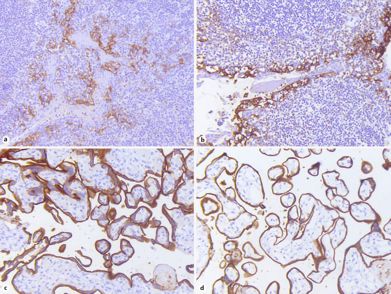
Fig. 2.
a–d PD-L1 IHC in FF versus CF tonsillar and placental tissue. Tonsillar (a, b) and placental control tissues (c, d) after direct formalin fixation (a, c) and CF (b, d) show similar PD-L1 immunohistochemical reactivity with minimal differences in staining quality (×200). PD-L1, programmed death ligand-1; IHC, immunohistochemistry; FF, formalin-fixed; CF, CytoLyt®-prefixed.

