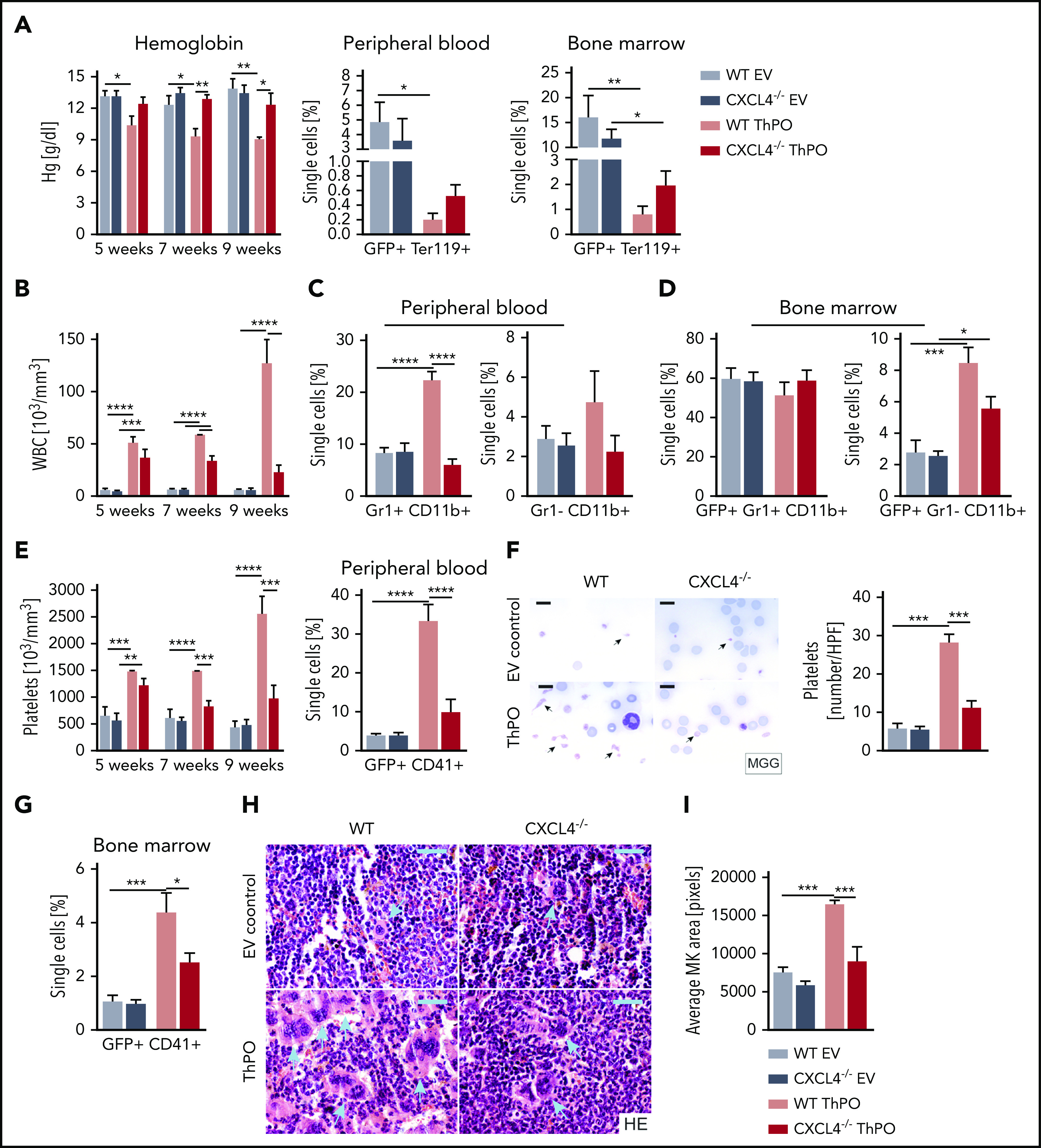
Figure 3.

Loss of Cxcl4 in hematopoietic cells ameliorates the MPN phenotype and restores MK and platelet abnormalities. (A) Hemoglobin (Hg) counts monitored over time, together with the frequency of GFP+ Ter119+ cells in peripheral blood and BM 9 weeks’ posttransplant (euthanasia). (B) WBC counts are monitored over time from peripheral blood. (C) Flow cytometric quantification of Gr1+ CD11b+ (granulocytes) and Gr1– CD11b+ (monocytes) at euthanasia in peripheral blood. (D) Flow cytometric quantification of GFP+ Gr1+ CD11b+ (granulocytes) and GFP+ Gr1– CD11b+ (monocytes) at euthanasia in BM. (E) Platelet counts are monitored at 5, 7, and 9 weeks’ posttransplant. Flow cytometric quantification of GFP+ CD41+ cells in peripheral blood. (F) May-Grünwald-Giemsa (MGG) staining and quantification of the number of platelets per high-powered field in murine blood smears. Original magnification ×100. Scale bar, 10 μm. (G) Flow cytometric quantification of GFP+ CD41+ (MKs) in BM. (H) Hematoxylin and eosin (HE) staining of 4-μm BM sections (femur) with particular focus on the size and morphology of MKs (blue arrows). Original magnification ×40. Scale bar, 50 μm. (I) Quantification of the mean area of MKs in BM. n = 5 mice/group, 3 males. Data are shown as mean ± standard error of the mean, 1-way analysis of variance followed by Tukey’s post hoc test. *P < .05, **P < .01, ***P < .001, ****P < .0001.
