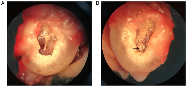
Figure 1.
Otomicroscopic observations at week 8. (A) Representative left ear used as the control. The tympanic membrane was intact and transparent without calcification. (B) Representative right ear used as the experimental model. The tympanic membrane showed obvious turbidity and sclerotic plaques (arrow) at the tympanic ring. Original magnification, x20.

