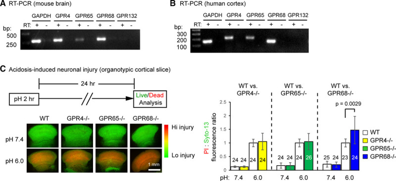
Figure 1.

Functional screening identifies GPR68 as a regulator of pH 6-induced neuronal injury. A and B, Reverse transcription (RT)-polymerase chain reaction (PCR) result on the expression of proton-sensitive GPCRs (G protein-coupled receptor) in mouse brain (A) and human cortical tissue (B). −RT has no reverse transcriptase added into RT. Images shown were from products of 35 cycles of PCR. C, Representative fluorescence images and quantification showing pH 6-induced neuronal injury in organotypic cortical slices from wild-type (WT) and corresponding GPCR knockouts. Organotypic cortical slices were treated with pH medium buffered at pH 7.4 or 6.0 for 2 h and stained for propidium iodide (PI, red) and Syto-13 (green) 24 h later. Relative fluorescence intensity of PI and Syto-13 was quantified. Increased red/green ratio indicates increased neuronal injury. Dashed lines on the bar graph indicate that the WT controls for the 3 sets were different. For all experiments, the knockouts were compared with the WT that was cultured and treated in parallel. To better compare different experiments, injury (PI:Syto-13 ratio) in pH 6 of WT in a given experiment was normalized to 1. N on the bars indicate total number of slices quantified from 6 different experiments.
