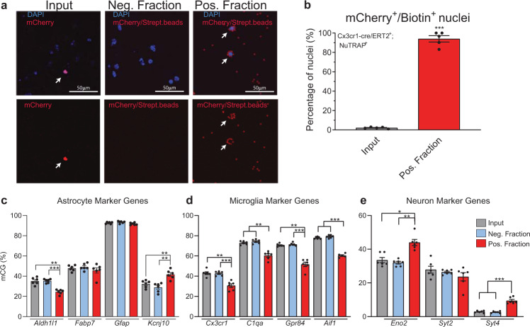
Fig. 8. Validation of microglial epigenome enrichment in the Cx3cr1-NuTRAP mouse brain by INTACT-BSAS.
a Representative confocal fluorescent microscopy images from input, negative, and positive INTACT nuclei fractions. Scale bar: 50 µm. b Purity of microglial nuclei expressed as average percentage ± SEM mCherry+/ Biotin+ nuclei in the positive fraction, and percentage ± SEM mCherry+ nuclei in the input and average percentage ± SEM mCherry+/Biotin+ nuclei in the input demonstrates a high degree of specificity to the INTACT isolation (n = 5/group, ***p < 0.001 by paired t-test comparing positive fraction to input). c–e INTACT-isolated genomic DNA from Cx3cr1- NuTRAP mice was bisulfite converted and DNA methylation in specific regions of interest (promoters for neuron, astrocytes and microglia marker genes) were analyzed by Bisulfite Amplicon Sequencing (BSAS) from input, negative, and positive fractions. Hypomethylation of the microglial marker genes Cx3cr1, C1qa, Gpr84, and Aif1 in the positive fraction compared to input and negative fraction was observed. Hypermethylation of neuronal markers Eno2 and Syt4 and astrocyte marker Kcnj10 was observed (n = 6/group, average % mCG ±SEM, RM One-way ANOVA with Tukey’s post-hoc, *p < 0.05, **p < 0.01, ***p < 0.001).

