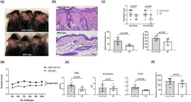
Figure 5.
Oxazolone-induced atopic dermatitis in HSD11B1 KO and WT mice. (a) Gross photographs of skin lesions in global HSD11B1 KO mice with AD induced by Ox and WT mice (n = 8/group) taken before sacrifice (at 26 days). (b) Macroscopic appearance of skin sections of dorsal lesional surface. Scale bar, 10 µm. (c) Histological scores for dermal inflammation and density measured as epidermal and dermal thickness. (d) Eczema scores for HSD11B1 KO and WT mice at challenges 5–10. (e) TEWL, SC pH and SC hydration measured before sacrifice (at 26th day). (f) Corticosterone levels in epidermis of KO and WT mice measured by ELISA (*p < 0.05, ** p < 0.005). Data are presented as means ± standard deviation. KO knock out, SC stratum corneum, TEWL transepidermal water loss.

