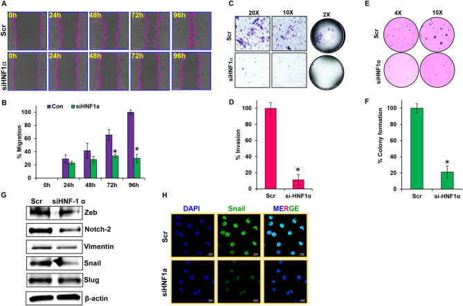
Figure 4.
HNF1α regulates metastatic characteristics of PDAC. (A,B) Wound-healing assay was performed in siHNF1α AsPC-1 cells; migration was analyzed using Nikon Biostation CT at 2 h intervals for up to 96 h at × 4 magnification. (C,D) Invasiveness of siHNF1α AsPC-1 cells and overexpression HNF1α HPAC cells were observed using a Matrigel invasion assay, and captured using Nikon Eclipse TS 100 microscope at × 20 and × 100 magnification. (E,F) Colony formation assay was performed in siHNF1α AsPC-1 cells. (G) EMT markers analyzed by western blot in siHNF1α AsPC-1 cells. (H) Immunofluorescence analysis of Snail in siHNF1α AsPC-1 cells, captured using Nikon SMZ 1500 microscope at × 40 magnification. Data shown as mean ± SEM. Experiments (n = 3) were repeated three times in triplicates. *p < 0.05.

