
Figure 4.
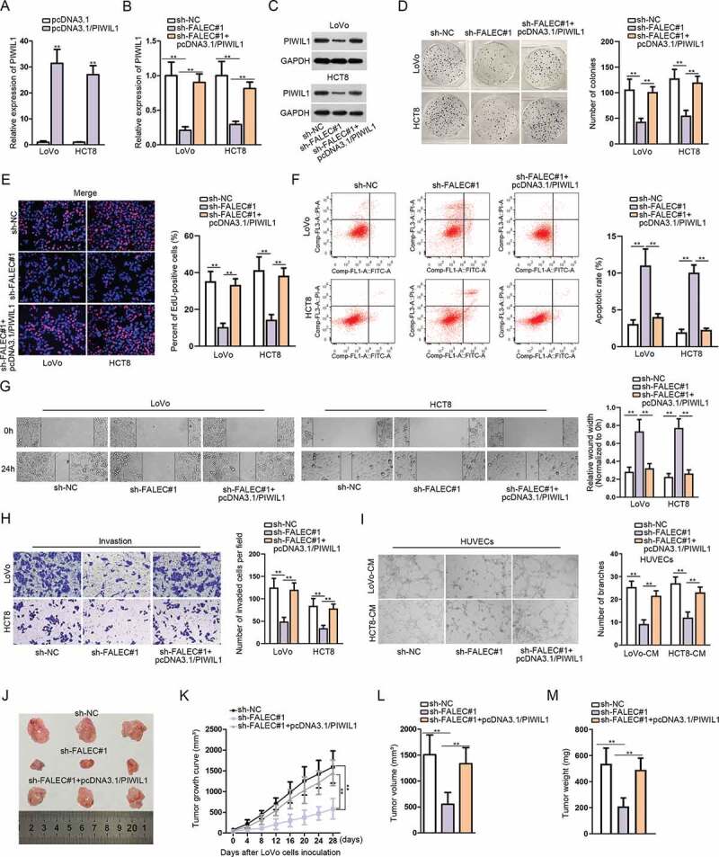
FALEC promotes CRC progression via targeting miR-2116-3p/PIWIL1 axis
A. The expression of PIWIL1 after transfecting with pcDNA3.1/PIWIL1 was detected by qRT-PCR analysis. B–C. The mRNA and protein levels of PIWIL1 after silencing FALEC were detected by qRT-PCR and western blot, respectively. D–E. Colony formation and EdU assays were performed to study the impact of overexpressing PIWIL1 on the proliferation of FALEC-silenced LoVo and HCT8 cells. F. Flow cytometry analysis was performed to assess the percent of apoptotic CRC cells under different conditions. G–I. Wound-healing, transwell and tube formation assays were performed to examine cell migration, invasion and angiogenesis under diverse contexts. J. Representative images of tumors excised from mice in sh-NC, sh-FALEC#1 or sh-FALEC#1+ pcDNA3.1/PIWIL1 group. K. The growth curve of above tumors recorded every 4 days during 4 weeks. L–M. Tumor volume and weight were measured and compared among sh-NC, sh-FALEC#1 and sh-FALEC1+ pcDNA3.1/PIWIL1 groups. **P < .01.