
Fig 6. Immunohistochemical analysis of PD-L1 in EMM.
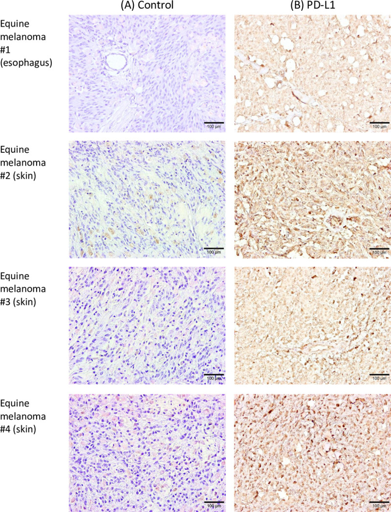
Immunohistochemical staining of PD-L1 in tumor tissues of horses with melanoma (#1–#4). Each section was stained (A) without a primary antibody (control) or (B) using anti-bovine PD-L1 mAb (6C11-3A11). Further information of tumor specimens is shown in S2 Table.