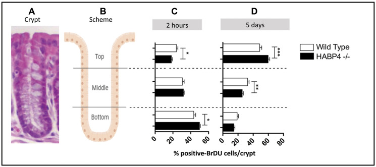
Figure 2. Increased proliferation in the colon epithelia of knockout mice.
(A) histological image of the overall crypt organization, (B) schematic representation of the three main regions analyzed. The BrdU incorporation assay was performed with the colons of the Habp4 –/– animals (black) and wild-type (white), after 2 hours (C) and 5 days (D) of the injection. The cells labeled with anti-BrdU were quantified in the three compartments of the crypt, by manual counting, and at least 20 crypts per animal were counted (number of mice: control 2 h n = 2; HABP4 –/– 2 h n = 5; control 5 days n = 3; HABP4 –/– 5 days n = 5). Data are presented as mean and SD. * p < 0.05, ** p < 0.005, *** p < 0.001.

