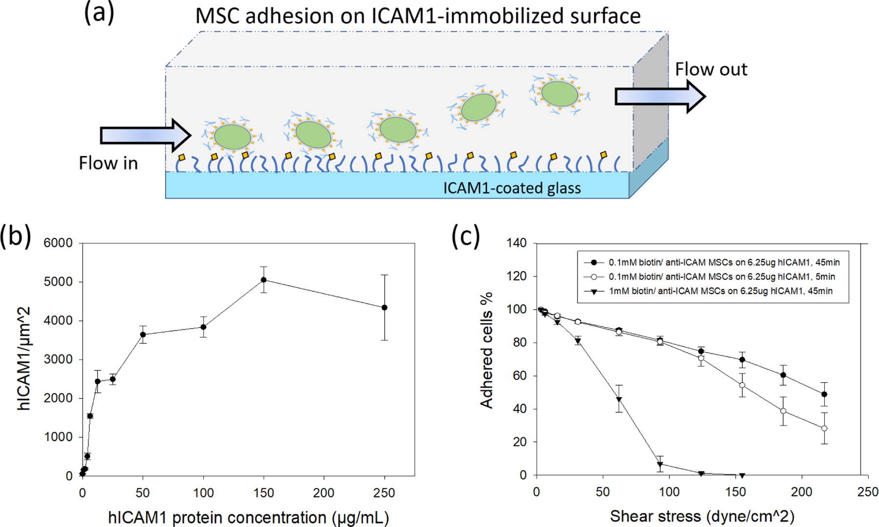
Figure 5. Cell adhesion on ICAM1-modifed glass and cell detachment under the presence of shear flow.

(a) Schematic diagram of experimental setup of shear-detached cells on ICAM1-modifed glass. (b) Relationship of human ICAM1 protein density and protein concentration used for epoxide glass modification. (c) Relationship between attached cells % and shear stress under different cell adhesion conditions (n=5 for 0.1mM biotin/anti-ICAM1 MSCs with 5 min; n=7 for 0.1mM biotin/anti-ICAM1 MSCs with 45 min; n=4 for 1mM biotin/anti-ICAM1 cells).
