. 2020 Nov 20;9:e61882. doi: 10.7554/eLife.61882

Figure 2. Reduced engram reactivation efficacy is correlated with impaired contextual fear memory in Fmr1 KO mice.

(A) Experimental protocol for activity-dependent genetic labeling of neural ensembles. Intensive fear conditioning training was used in constitutive Fmr1 KO mice to facilitate labeling. (B) Representative images showing memory-encoding neural ensembles (engram cells) labeled with tdTomato (red) and memory recall-activated neurons labeled with FOS immunostaining (green). The circles in zoomed in images highlight reactivated neurons (yellow). Scale bar: 100 μm. (C) Quantification of percentage of neurons activated during learning. (D) Quantification of percentage of neurons activated during memory recall. (E) Quantification of engram reactivation efficacy in CA1 as percent FOS-positive neurons in tdTomato-positive and tdTomato-negative populations [one-way ANOVA with Tukey’s multiple comparison test: F (3, 32)=26.57, p<0.0001, ***p<0.001; **p<0.01]. (F) Positive correlation between neural ensemble reactivation efficacy and behavioral perform during context memory test (**p<0.01, Pearson correlation coefficient). n/N, number of mice/number of independent litters. All graphs represent mean ± SEM.

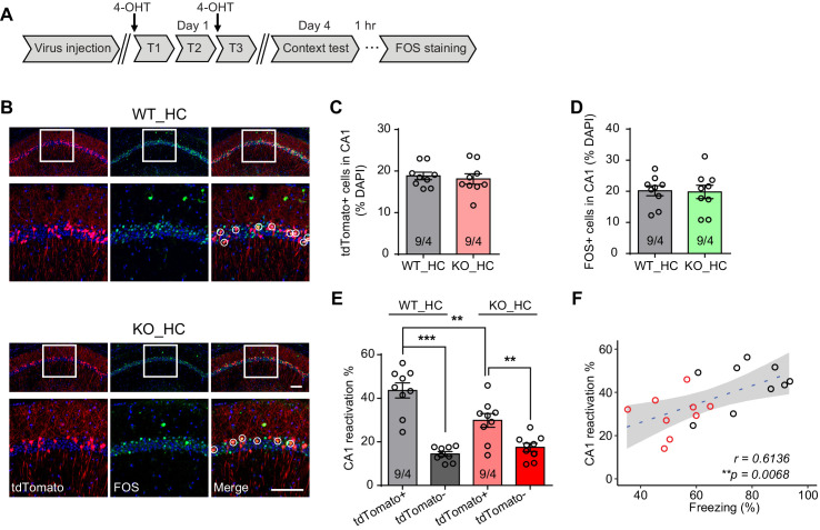
Figure 2.

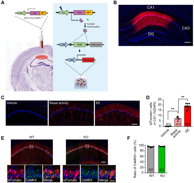
Figure 2—figure supplement 1. Validation of activity-dependent genetic labeling method and characterization of Fos-TRAP-labeled neurons.

Figure 2—figure supplement 1.

(A) Schematics of AAV-Fos-TRAP application. Ai9 reporter mice were injected with AAV-Fos-ERT2-Cre-ERT2-PEST at hippocampal CA1. Behavioral experience (fear conditioning training or enriched environment) activates Fos expression, which drives Cre expression and Cre-mediated recombination in the presence of tamoxifen, labeling a population of activated cells (tdTomato+) in the CA1. Non-active cells do not undergo recombination due to lack of Cre expression. (B) A representative image of a hippocampal tissue section showing intensive fear conditioning training-activated neurons in CA1. Scale bar: 200 μm. (C) Representative images showing the expression of tdTomato in CA1 after mice underwent 4 hr enriched environment. Scale bar: 100 μm. Mice with vehicle injection (Vehicle) or maintained in home cage with basal activity were used as controls. (D) Quantification of activated cells (tdTomato+) in CA1 [Kruskal-Wallis test with Dunn’s multiple comparison test: H (2)=15.41, p<0.001, **p<0.01]. (E) Representative images showing Fos-TRAP-tdTomato expression and CaMKII immunostaining in wild type and Fmr1 KO mice. Ai9 Mice expressing tdTomato reporter were injected with Fos-TRAP viruses in hippocampal CA1 region and trained with intensive fear conditioning. Learning activated neurons were labeled by tdTomato expression. Excitatory principle neurons in the hippocampus were identified with CaMKII immunostaining. Scale bars: 100 μm (upper) and 20 μm (lower). (F) Quantification of CaMKII-positive neurons in the tdTomato-expressing population. n/N, number of mice/number of independent litters. All graphs represent mean ± SEM.