
Fig. 1. KSHV ORF33 inhibits STING- and MAVS-mediated IFNβ production.
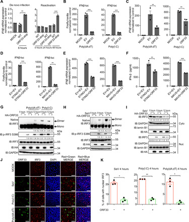
(A) IFNβ mRNA levels during de novo infection and reactivation of ORF33-null KSHV. THP-1 cells were infected with WT or ORF33-null KSHV at 50 genome copies per cell for 6 hours (left). iSLK cells carrying WT KSHV BAC or ORF33-null KSHV BAC were induced for 24 or 48 hours (right). (B to F) Influence of ORF33 on IFNβ production. Human embryonic kidney (HEK) 293 cells were transfected with the indicated expression plasmids (B to F), along with IFNβ-luc and TK-Renilla reporter plasmids (B and D), for 24 hours. Cells were challenged with transfection of poly(dA:dT) (1 μg/ml) (left) or poly(I:C) (1 μg/ml) (right) for 18 hours (B) or for 12 hours (C). Luciferase assays (B and D), RT-qPCR (C and E), and ELISA (F) were conducted. (G to K) Impact of ORF33 on dimerization, phosphorylation, and translocation of IRF3. HEK293 cells were transfected with the indicated expression plasmids for 24 hours. Cells were challenged with poly(dA:dT) or poly(I:C) (1 μg/ml each) or SeV infection (50 HA U/ml) for 12 hours (G to I) or 4 hours (J and K). (G and H) p-IRF3 levels were measured by SDS–polyacrylamide gel electrophoresis (PAGE), and IRF3 dimerization levels were measured by native-PAGE and quantified using ImageJ and normalized to IRF3 and glyceraldehyde-3-phosphate dehydrogenase (GAPDH). (I) Nuclear (Nuc) and cytoplasmic (Cyto) fractions were separated, and IRF3 levels were quantified using ImageJ and normalized to lamin B1 or GAPDH. (J) Cells were fixed and immunostained with anti-HA and anti-IRF3 antibodies. Scale bars, 40 μm. DAPI, 4′,6-diamidino-2-phenylindole. (K) The percentage of cells with nuclear IRF3 in ORF33-expressing or -nonexpressing population under the three different stimulations was calculated. (A to F and K) Data presented are means ± SEM of three independent measurements, representative of three independent experiments. *P < 0.05, **P < 0.01, and ***P < 0.001; Student’s t test, n = 3. See also figs. S1 to S3.