
Fig. 2. KSHV ORF33 impairs the recruitment of IRF3 by STING and MAVS.
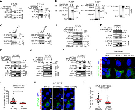
(A to C) Interaction between ORF33 and STING/MAVS. (A) HEK293T cells were transfected with indicated expression plasmids for 36 hours before coimmunoprecipitation. (B) GST-ORF33-His proteins were used to pull down lysates of HEK293T cells expressing HA-STING/HA-MAVS. (C) iSLK.219 cells were induced for 48 hours, and antibodies to STING and MAVS were used to enrich endogenous STING and MAVS, respectively. KSHV ORF33 protein was detected by an anti-ORF33 antibody. IgG, immunoglobulin G. (D to H) Impact of ORF33 on the key molecular events of the IFNβ production pathway. HEK293 cells were transfected with the indicated expression plasmids for 20 hours before coimmunoprecipitation. The gray values are quantified using ImageJ. (I to L) Analysis of the impact of ORF33 on the interaction between STING/MAVS and IRF3 in situ by PLA. HEK293T cells were transfected with the indicated expression plasmids: (I) MYC-ORF33, HA-IRF3, and green fluorescent protein (GFP)–STING or EV control(s); (K) MYC-ORF33, HA-IRF3(2A), and GFP-MAVS or EV control(s). Scale bars, 2.5 μm (I) and 10 μm (K). (J and L) The number of red fluorescent dots in each cell in (I) and (K) was enumerated, respectively. (J and L) Data presented are means ± SEM of three independent measurements, representative of three independent experiments. ***P < 0.001, Student’s t test. See also fig. S4.