
Fig. 1. Design of HNCIB system.
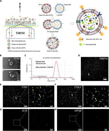
(A) Illustration of the HNCIB system for simultaneous detection of PD-L1 membrane protein and mRNA in a single EV. Photo credit: Ming Wang, Hangzhou Dixiang Co. Ltd., Hangzhou, China. (B) Electron micrograph showing the EVs isolated from human plasma. (C) Particle size and zeta potential measurement of the EVs isolated from human plasma by nanoparticle tracking analysis technique. d.nm, diameter (nm). (D) Bright-field TIRFM images showing the EVs isolated from human plasma. (E) EV marker CD9 and CD63 (F) membrane protein measured by the HNCIB system. (G) Non-EV markers ALB and APOB (H) protein measured by the HNCIB system.