
Fig. 3. Double immunofluorescence staining of CD63 and PD-L1 membrane protein.
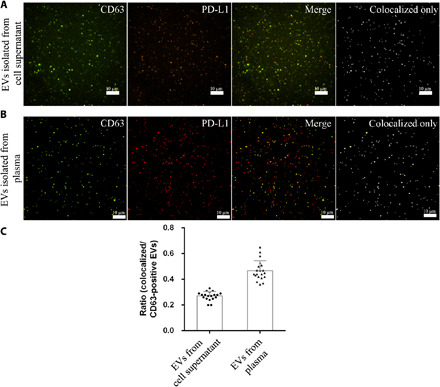
(A) EVs isolated from 1000 μl of cell supernatant of human A549 cells overexpressed with CD63-GFP and PD-L1–mCherry. The dots in the colocalized only image represent EVs expressing both CD63 and PD-L1 protein. (B) EVs isolated from 90 μl of human plasma are stained with CD63-AF488 and PD-L1–AF647 antibodies. The dots in the colocalized only image represent EVs expressing both CD63 and PD-L1 protein. (C) Ratio of CD63- and PD-L1–positive EVs to CD63-positive EVs isolated from human A549 cells overexpressed with CD63-GFP and PD-L1–mCherry or isolated from plasma of 20 patients with LUAD.