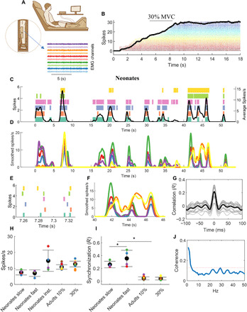
Fig. 4. Estimates of motoneuron output for the adult and neonatal human spinal cord.

(A) Twelve adults performed isometric ankle-dorsiflexion contractions at 10 and 30% of maximal force for 50 s. (B) The experimental setup and a representative isometric contraction with the corresponding motor unit activity. (C) Motor unit spike raster and after convolution with a 1-s Hann window [smoothed spikes per second (D)] for a representative newborn during spontaneous movements. The black line in (C) represents the average discharge frequency calculated after convolving the sum of the spike timings. Note the highly synchronized discharge timings in the scaled version (E and F). The cross-correlation value from the smoothed discharge timings was 0.52. (G) The cross-correlation function for 50 permutations of two equally sized groups of motoneurons (gray lines) and the corresponding average (black line). (H) The average motor unit discharge rate of the neonate’s individual movements and for the adults during the two tasks. The discharge rate was averaged in the intervals corresponding to slow or fast neonatal movements. Moreover, the average value of the instantaneous discharge rate across the full duration of the tasks (neonate inst.) is also reported. (I) The average motoneuron synchronization for each neonate and the adults. The neonates showed a higher synchronization value compared to the adults during both slow and fast movements (*P < 0.001). Moreover, the neonates exhibited higher synchronous firing during fast movements. (J) The average coherence across all neonates. The dashed red line indicates the significance threshold.
