Fig. 1.
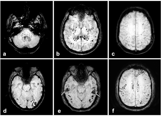
Susceptibility-weighted MR images in four different critically ill COVID-19 patients: a–c: diffuse microhemorrhages in a 66-year-old man appearing as multiple small hypointense foci within the brainstem and cerebellum (a), the splenium of the corpus callosum (arrowheads) and the internal capsules (arrows) (b), the juxtacortical white matter (c). d, e: Juxtacortical hematomas (arrows) associated with diffuse microhemorrhages in men of 60 (d) and 67 years (e). f subarachnoid hemorrhages (arrows) associated with diffuse microhemorrhages in a 57-year-old man.

