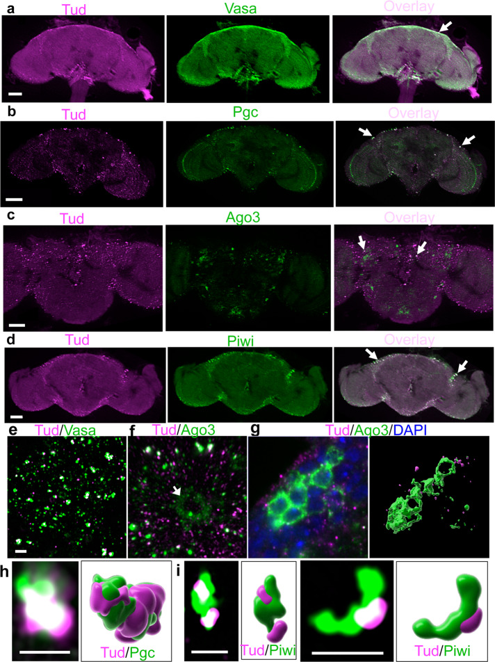
Fig. 3. Vasa, Polar granule component, Ago3, and Piwi colocalize with Tudor in glial granules.
a–d Optical sections of the adult brains immunostained with anti-FLAG antibody to label Tud (magenta) and Vasa (a), Pgc (b), Ago3 (c), and Piwi (d) (green) show the localization of all these proteins in glia. Arrows point to colocalized foci. e, f Super-resolution images with Tud and Vas (e) and Ago3 (f, Supplementary Movie 1) glial granules. In addition to glia, Ago3 is expressed in neurons which do not express Tud (a neuronal cell body is indicated with an arrow (f)). g In neurons, Ago3 is frequently expressed in the cytoplasm of several neuronal cell bodies clustered in the cortex glia (green) with a characteristic “honeycomb” appearance (super-resolution optical section, left panel). Right panel shows 3D reconstruction of the Ago3-positive neurons (green) and Tud glial granules (magenta) corresponding to the left image. h, i Super-resolution optical sections and corresponding 3D reconstructions of Tud/Pgc (h, Supplementary Movie 2) and Tud/Piwi (i) individual glial granules (Tud and Pgc/Piwi are labeled with magenta and green respectively). Scale bar in a, b, d, and c is 50 μm and 40 μm, respectively; 2 μm scale bar in e is the same for f and g. Scale bar in h and i is 2 μm.

