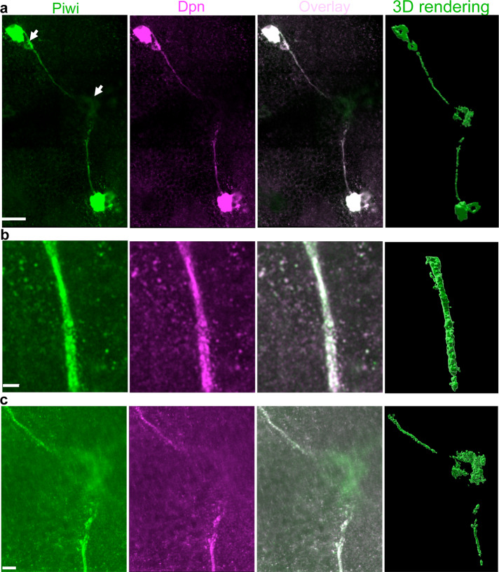
Fig. 5. Piwi/Deadpan-expressing cells form long extensions in adult brains.
a Super-resolution imaging of Piwi (green channel) and Dpn (magenta)-positive extensions, which emanate from the cells and end in the brain midline converging to a Piwi-positive cell (indicated with arrow at the middle of the left panel). Also, a smaller Piwi/Dpn+ cell appears to be connected to a larger neighboring cell by the extension (indicated with top arrow). b A super-resolution optical section showing details of an extension’s segment. c A super-resolution optical section of the brain midline showing details of the central segments of the extensions converging in the Piwi-positive cell. Right panels in a–c show the corresponding 3D reconstructions based on super-resolution optical sections (green channel). To visualize extensions, the images were generated with either high laser power during acquisition or post-acquisition by increasing the signal intensity with Imaris software. Scale bars in a, b, and c are 20, 2, and 5 μm respectively.

