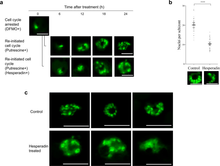
Fig. 2. Nuclei number and aberrant nuclear morphology of parasites after Hesperadin treatment.
Intraerythrocytic P. falciparum 3D7 parasites were synchronized with DFMO and then allowed to re-initiate their cell cycle by addition of putrescine, in the presence or absence of Hesperadin (24 h treatment). a Parasites were sampled every 6 h after hesperadin treatment and life cycle progression monitored through SYBR Green I fluorescence microscopy using a Zeiss LSM 880 Confocal Laser Scanning Microscope (LSM). b Control and hesperadin-treated parasite nuclei development was quantitatively assessed through SYBR Green I fluorescence microscopy. Inter-nuclei distances were determined for a minimum of ten nuclei. Significant difference was calculated using two-tailed equal variance Students t-test, ****P < 0.0001, n = 36. Error bars represent 95% CI of the mean. Scatter plots were generated using GraphPad Prism version 6.01 software. Data are available in Supplementary Data 1. c Representative pictures of morphological abnormalities observed in Hesperadin-treated nuclei for three individual schizonts each. Scale bars apply to all micrographs and correspond to 5 µm.

