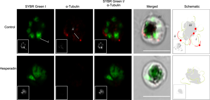
Fig. 4. Relative positions of SYBR green and AlexaFluor 647-conjugated anti-α-tubulin-stained material in untreated (control) and Hesperadin-treated parasites.
The column labelled ‘Merged’ is a superposition of the fluorescence images in the red and green channels with a differential interference contrast image of the same parasite. The column labelled ‘Schematic’ is an interpretation of the relative positions of the nuclear material (i), the mitotic spindle (ii) and haemozoin crystals in the food vacuole (iii); actual images of nuclear and spindle material are also indicated for a single daughter cell in the control row. Images are representative of at least seven parasites evaluated per sample. Black and white versions of the colour pictures have been added as inserts. Scale bars apply to all colour micrographs and correspond to 5 µm.

