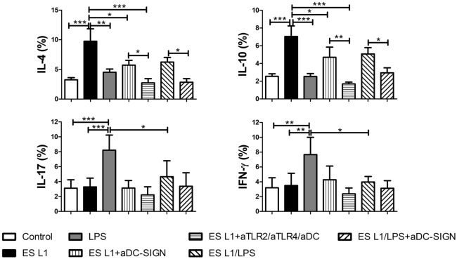
Figure 6.
Impact of DC-SIGN on T helper polarization induced by ES L1-pulsed dendritic cells (DCs). DCs treated with specific DC-SIGN blocking antibodies, alone or simultaneously with TLR2 and TLR4 blocking antibodies, prior to ES L1 and/or LPS, were washed thoroughly and then cocultured with magnetic-activated cell sorting-purified allogenic T cells (Tly) (1 × 10⁵/well) for 6 days in 1:20 DC:T cell ratio. Upon cocultivation, the percentage of cytokines expression was measured intracellularly by flow cytometry, within the T cells subjected to CD4 surface staining prior to intracellular staining, and treated with PMA/Ionophore/monensin for the last 4 h. The summarized results are shown as mean percentages (%) ± SD of three experiments with different DCs donors. *p < 0.05, **p < 0.01, ***p < 0.001 compared to control (non-treated cells cultivated in medium), or as indicated (one-way ANOVA with Tukey’s posttest).

