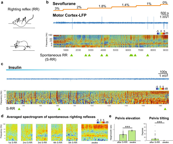
Figure 1.
Spontaneous righting reflex is associated with low arousal state. (a) Schematic of spontaneous righting reflex. The animal is placed on its back and the subject rocks the trunk to the right and left side together with stretching of head and limbs. This results in rotation of the body so that all four limbs touch the ground (b) Representative trace of motor cortex raw LFP and normalized spectrogram during emergence from sevoflurane anesthesia. Color bar shows power in decibels. Light green triangles represent time points at which spontaneous righting reflexes (S-RR) were observed while the animal whose spectrogram is shown in the figure emerged from anesthesia and (c) hypoglycemic coma induced by injecting insulin (d) Average cortical spectrogram (60 s) at a time when the first five spontaneous righting reflexes occurred in animals exposed to anesthetic and insulin (n = 13 animals). Data was compared to the averaged spectrogram (120 s) obtained from the same group of animals once they regained full motor activity and wakefulness. (e) Quantification of pelvis elevation and tilting of the hips as an indirect measure of erect posture after S-RR. We measured the height/width ratio and the tilting angle of an ellipse that contours the animal hip and limbs (n = 52 S-RR; p = 0.0001 and p = 0.0001 respectively; Mann–Whitney U test and Two-Sample Proportion Test; See methods). ***p = 0.001.

