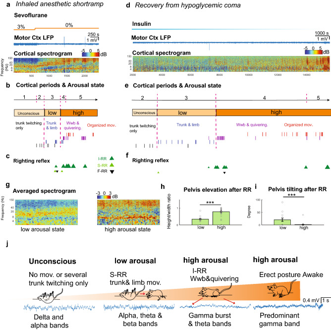
Figure 3.
Integration of cortico-motor activity accurately determines the level of arousal. (a) Representative trace of LFP recorded in motor-cortex and normalized spectrogram during a short ramp of sevoflurane. Color bar represents power in decibels. (b) Segmented cortical periods and progression of motor behavior restoration defined high and low arousal states in the subject. (c) Distinct RR events including induced RR (I-RR), spontaneous RR (S-RR) and failed RR after perturbation (F-RR) during emergence from an animal exposed to a short ramp of anesthetic. (d) Motor cortex LFP and spectrogram of an animal recovering from hypoglycemic coma. (e) Segmented cortical periods and progression of motor behavior restoration define high and low arousal states in the hypoglycemic mouse (f) Dissimilar RR events observed during restoration of an awake state in an animal injected with insulin (g) Averaged spectrogram (1000 s) of motor cortical activity during a low (n = 5) and high (n = 5) arousal state. Color bar represents power in decibels. (h) Quantification of pelvis elevation and (i) tilting after RR in low (n = 17) and high arousal (n = 30); p < = 0.001 and p = 0.0005; Mann–Whitney U test and Two-sample Proportion Test). ***p = 0.001. (j) Schematic illustrates levels of arousal defined by cortico-motor features during restoration of an awake state.

