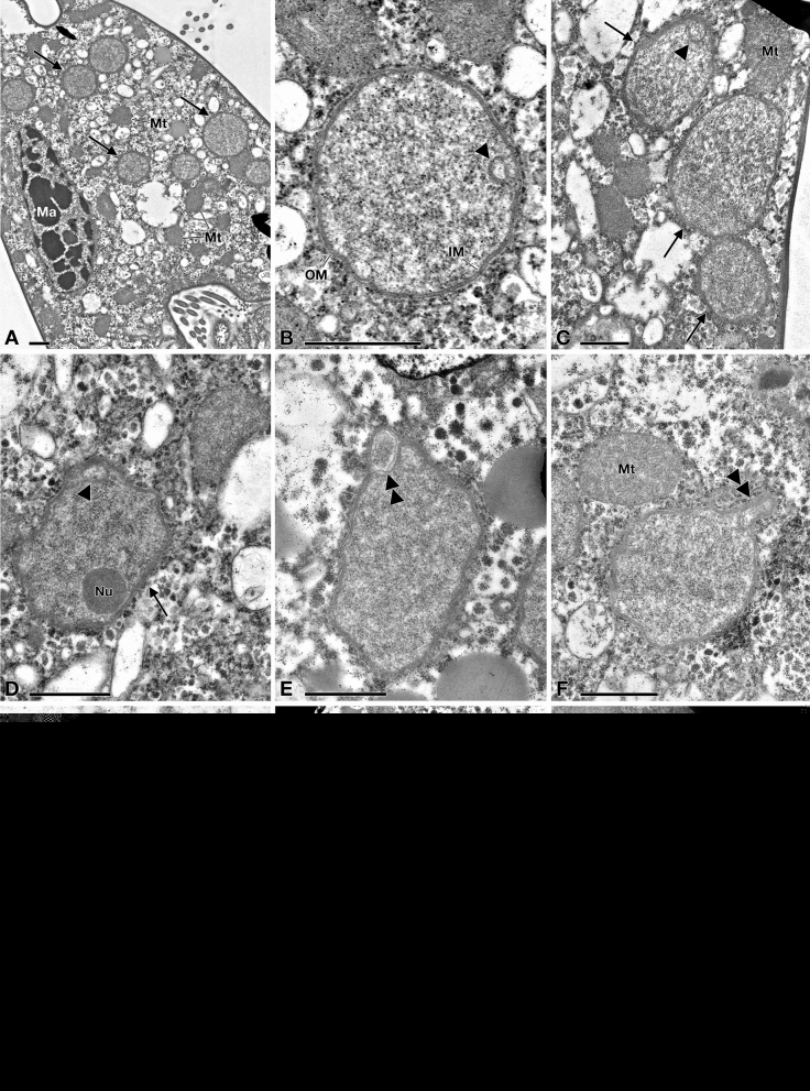
Figure 5.
TEM pictures of "Candidatus Pinguicoccus supinus". (A) Endosymbiont cells (arrow) in host cytoplasm, lying underneath the cortex, aggregated in clusters; macronucleus (Ma) and some mitochondria (Mt), also in proximity or in apparent close contact with endosymbionts, are visible; (B) Closer view of “Ca. Pinguicoccus supinus” showing inner membrane (IM) and outer membrane (OM); an invagination of the inner membrane (IM) is present (arrowhead); (C) Different cell shapes of three “Ca. Pinguicoccus supinus” specimens (arrow), from rounded to ovoid; the IM is invaginated (arrowhead); (D) Endosymbiont cell with an irregular shape, with an evident nucleoid (Nu) and an invagination of the IM is present (arrowhead); (E,F) “Ca. Pinguicoccus supinus” cells showing evagination of the outer membrane (double arrowhead); (F) Endosymbiont cell in proximity to a mitochondrion; (G) “Ca. Pinguicoccus supinus” showing emphasized folding (asterisk) of membrane area; (H) Endosymbiont cells (arrow) appear to be in close contact with lipid droplets (L); (I) “Ca. Pinguicoccus supinus” (arrow) during binary fission; the division septum is well visible. IM inner membrane, L lipid droplet, Ma macronucleus, Mt mitochondrion, Nu nucleoid, OM outer membrane. Arrow: “Ca. Pinguicoccus supinus” cell; Arrowhead: evagination of the outer membrane; Asterisk: folding of membrane area; Double arrowhead: section of bacterial cell folding. Bars stand for 1 µm (A–F,H,I) and 0.5 µm (G).

