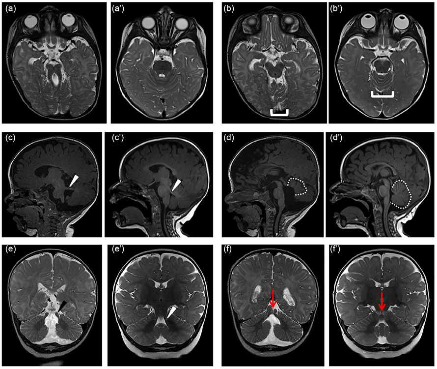
FIGURE 1.
Diagnostic features of Joubert syndrome on magnetic resonance imaging (MRI). (a) molar tooth sign (MTS) on T2-weighted axial images due to the combination of long, thick superior cerebellar peduncles (the roots of the tooth) and a deep interpeduncular fossa (the cutting surface of the tooth). This appearance is not always captured in a single plane, so it is important to evaluate the MRI as a whole rather than relying on a single image. (b) Superior cerebellar foliar dysplasia (bracket) on axial T2-weighted images, sometimes seen in the absence of the MTS in individuals carrying pathogenic variants in the JS-associated genes (note that the folia at this level are usually shaped like a U or V). (c) Horizontally oriented superior cerebellar peduncle (white arrowhead) on T1-weighted parasagittal image; (d) Vermis hypoplasia (outline), elevated roof of the fourth ventricle, and rostrally displaced fastigium on T1-weighted sagittal image; (e) Long, thick superior cerebellar peduncles (black arrowhead) T2-weighted coronal image; (f) Appearance of a cerebellar cleft (black arrow indicating fluid at the cerebellar midline) on T2-weighted coronal image. (a'-f') similar images from an unaffected individual of the same age

