
FIGURE 5.
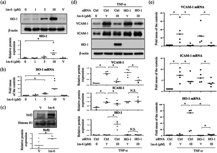
1m‐6 blocks TNF‐α‐induced intercellular adhesion molecule 1 (ICAM‐1) expression by inducing HO‐1 expression in human umbilical vein endothelial cells (HUVECs). (a and b) HUVECs were treated with various doses of 1m‐6 for 24 h. (a) Cell lysates were collected and analysed using western blot. Representative data and quantitative results expressed as the median with individual data of five independent experiments are shown. (b) Supernatants were collected and analysed using elisas. Data are shown as the median with individual data of five independent experiments. (c) HUVECs were treated with 10‐μM 1m‐6 for 24 h. Nuclear lysates were collected and analysed using western blot. Representative data and quantitative results of five independent experiments expressed as the median with individual data are shown. (d and e) HUVECs were transiently transfected with siCtrl or siHO‐1 for 24 h. After transfection, the cells were treated with 10‐μM 1m‐6 or DMSO for 2 h and further stimulated with 10 ng·ml−1 TNF‐α for 22 h. (d) Cell lysates were collected and analysed using western blot. Representative data and quantitative results expressed as the median with individual data of five independent experiments are shown. (e) Cellular mRNA was collected for further qRT‐PCR analysis. Data are shown as the median with individual data of five independent experiments. V indicates the vehicle (DMSO) control. Significance is presented as * P < 0.05 versus the indicated group. N.S. indicates non‐significant