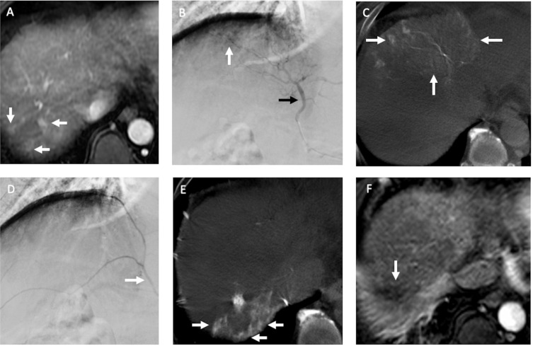
Figure 2.
Recurrent HCCs in a 60-year old man with nonB and nonC cirrhosis of liver and HCC in segment seven received radiofrequency ablation (RFA) before. (A) The arterial phase of dynamic MRI study before TACE showed three small recurrent HCCs (arrows) in segment seven of the liver abutting prior RFA site. (B) During TACE, the DSA of middle hepatic artery (black arrow) demonstrated suspected hypervascular tumor stains (arrow) near liver dome. (C) However, the CBCT-HA of middle hepatic artery indicated it supplied segments four and eight of the liver (arrows), not segment seven where recurrent HCCs were located. (D) The DSA of right inferior phrenic artery (RIPA) (arrow) revealed no distinct hypervascular tumor stain. (E) The following CBCT-A of RIPA confirmed it supplied the recurrent HCCs (arrows) in segment seven of the liver, and chemoembolization with drug-eluting beads was then performed. (F) One month after TACE, the MRI study indicated complete response (arrow) of the recurrent HCCs.

