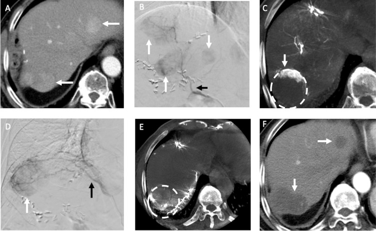
Figure 3.
Recurrent HCCs in an 83-year-old man with HBV cirrhosis of liver and HCC in segment seven received hepatectomy before. (A) The arterial phase of MDCT before TACE showed two recurrent HCCs (arrows) in the both lobes of liver. (B) During TACE, the DSA of right hepatic artery (black arrow) demonstrated three hypervascular tumors (arrows) in both lobes of the liver (one was not in the same cut of MDCT). (C) The following CBCT-HA of right hepatic artery indicated the recurrent HCC in segment seven (dotted line) was only enhanced in its anterior portion (arrow); the nonenhanced portion may be supplied by other vessels. (D) The DSA of RIPA (black arrow) showed a hypervascular tumor (arrow) near liver dome. (E) The following CBCT-A of RIPA confirmed the posterior portion of recurrent HCC in segment seven (dotted line) was supplied by RIPA, and chemoembolization with drug-eluting beads was then performed. (F) One month after TACE, the MDCT indicated complete response (arrows) of the recurrent HCCs.

