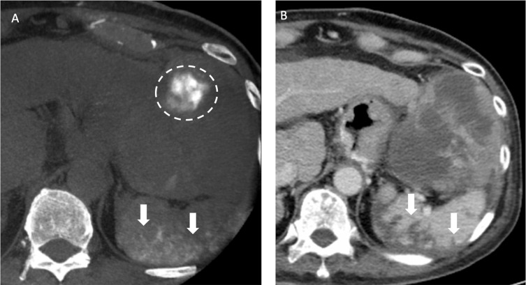
Figure 7.
Recurrent HCCs in a 70-year old man with HBV cirrhosis of liver and HCC in the both lobes of the liver received TACE before. (A) After the chemoembolization of splenic artery with drug-eluting beads, the nonenhanced CBCT after TACE showed contrast retention in the spleen (arrows), indicative of nontarget embolization. Also note contrast retention in the HCC located in the left lateral segment of liver (dotted line). (B) The follow-up CT one month after TACE showed wedge-shaped poor-enhancing area (arrows) in the spleen on portal venous phase, compatible with splenic infarction caused by nontarget embolization during TACE.

