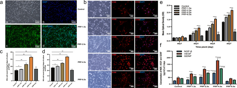
Fig. 1. The proliferation and neurotrophin expression of SCs treated with PRP.
a Immunostaining of harvested SCs. b SCs exposed to various PRP concentrations during 5 days of culture. c, d displayed the SCs count during 3 days and 5 days of culture (*P < 0.05, **P < 0.01). e CCK-8 colorimetric assay was performed to evaluate the effect of PRP in various concentration on the proliferation of SCs (**P < 0.01 vs. control group; ††P < 0.01 vs. PRP-6.5× group; ‡‡P < 0.05 vs. other five groups). f The NGF-β, VEGF and GDNF secreted by SCs that cultured with PRP at different concentrations were measured by ELISA at 5 days of culture (*P < 0.05 **P < 0.01 vs. control group; ††P < 0.01 vs. PRP-6.5× group; ‡‡P < 0.05 vs. other five groups). Error bars = SD, n = 3. SCs Schwann cells, PRP platelet-rich plasma, CCK-8 Cell Counting Kit-8.

