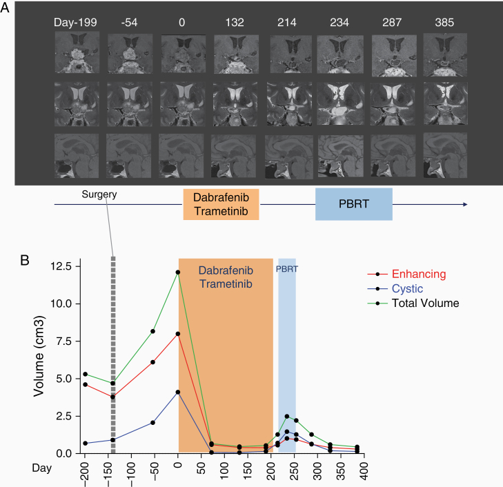
Figure 1.
Targeted inhibition of BRAF/MEK pathway in a patient with a recurrent papillary craniopharyngioma. (A) Timeline from the first presentation is indicated in days. T1-weighted (sagittal), contrast-enhanced (coronal) and T2-weighted (coronal) images of MRI performed at diagnosis (day –199). Follow-up MRI performed 3 months after surgery showing progression (day –54). Medical therapy with dabrafenib and trametinib was started on day 0. Subsequent MRI during medical therapy showed major tumor response on days 72 and 132. Anti-BRAF and anti-MEK inhibitors were stopped on day 208 and RT was started on day 215. After the anti-BRAF and anti-MEK discontinuation and during RT we observed an initial increase of the tumor, mainly of the cystic component (on days 214 and 234) followed by stabilization and final reduction (day 327, corresponding to the last follow-up). Tumor volume measure at corresponding time points is shown in (B). Enhancing (red) and cystic (blue) components as well as the total volume (green) variations are indicated. Tumor volumes were calculated by semiautomatic segmentation using Myrian Expert, v2.7.1, Intrasense, France.software. Of note, in MRI on day 0 contrast-enhancement is reduced (ie, half gadolinium dose compared to other MRIs) due to incidental extravasation during the contrast-agent infusion. PBRT, proton beam radiation therapy.

