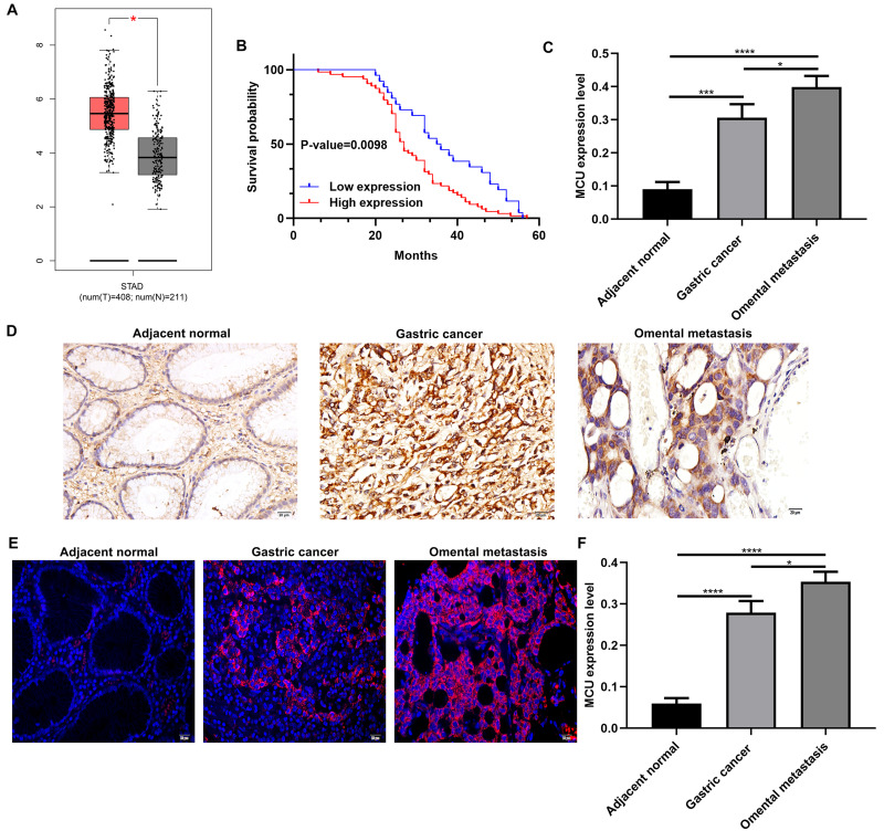
Figure 1.
MCU is significantly highly expressed in gastric cancer and associated with clinical characteristics and prognosis of patients with gastric cancer. (A) Comparison of MCU expression between gastric cancer tissues and normal tissues. (B) Kaplan–Meier survival analysis of correlation between MCU expression and gastric cancer prognosis in a cohort of 90 gastric cancer patients. Red represents high expression and blue represents low expression. (C) According to immunohistochemistry results, MCU had the higher expression levels in gastric cancer tissues or omental metastasis than adjacent normal tissues. (D) Representative images of immunohistochemistry results of MCU expression in 90 pairs of gastric cancer tissues and adjacent normal tissues. (E) Representative images of immunofluorescence of MCU expression in gastric cancer tissues and adjacent normal tissues. (F) High MCU expression was detected in gastric cancer tissues. Red fluorescence expresses MCU expression. *p<0.05; ***p<0.001; ****p<0.0001.

