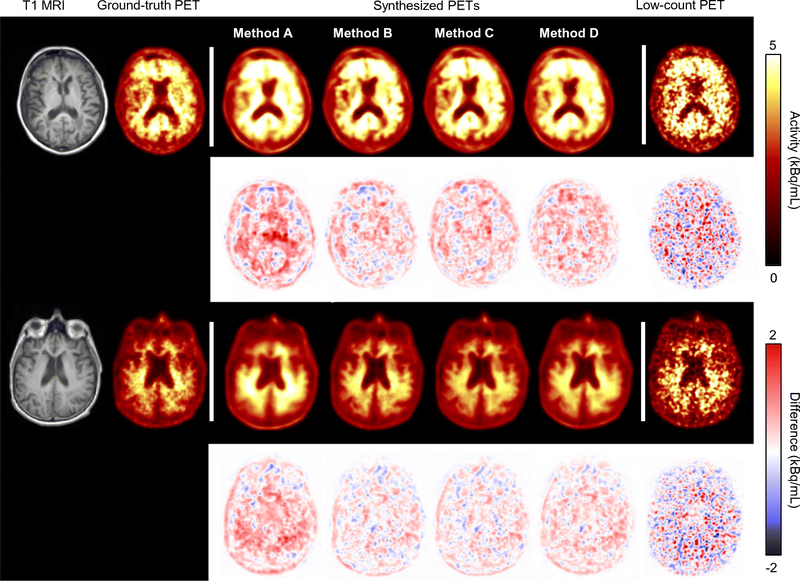
Figure 3.
Representative amyloid positive (top)/negative (bottom) images, with T1-weighted MRI and the corresponding PET images overlaid onto the T1 images. Difference images between the ground-truth and the other images are also shown. All synthesized images show marked noise reduction. However, Method A images are blurrier than the other synthesized images. Network training methods: A-direct application of pre-trained network; B-transfer learning starting with pre-trained network; C-training new network from scratch; D-training new network with combined datasets

