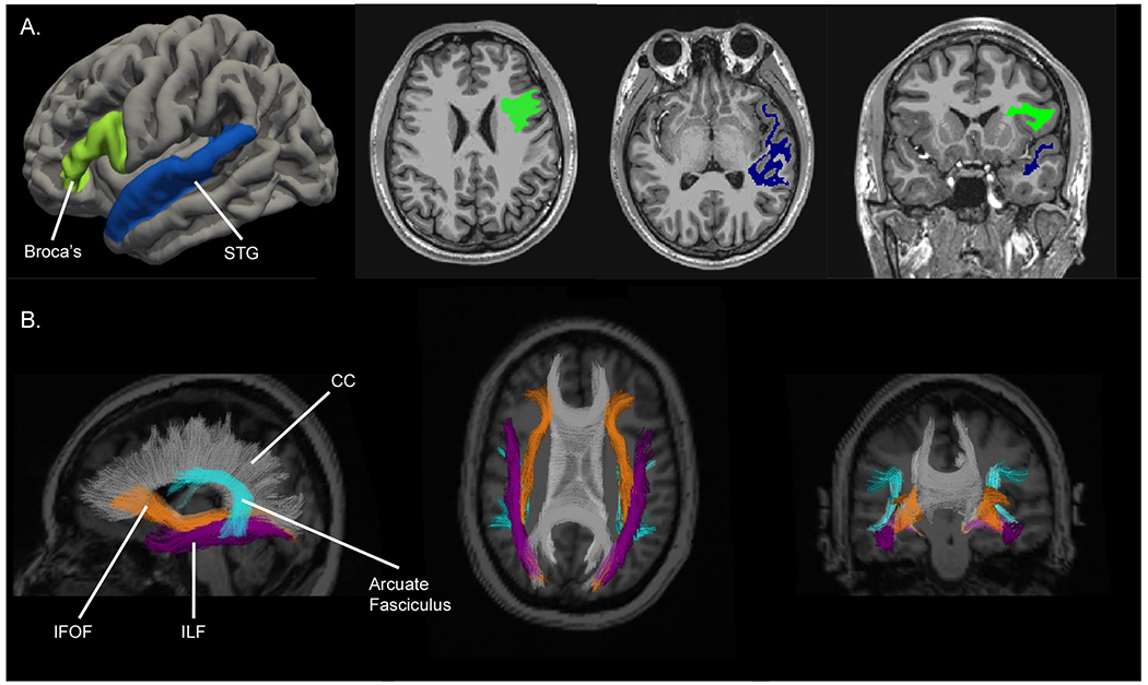
Figure 1.

Representative segmentation of ROIs overlaid on an MRI image. A) Left Broca’s Area (bright green) and left STG (navy blue) superficial WM ROIs. B) WM tracts of IFOF (orange), ILF (purple), and Arcuate fasciculus (turquoise) ROIs. The CC is segmented in gray and labeled for reference only.
Abbreviations: WM, white matter; ROIs, regions of interest; STG, superior temporal gyrus; IFOF, inferior fronto-occipital fasciculus; ILF, inferior longitudinal fasciculus; CC, corpus callosum.
