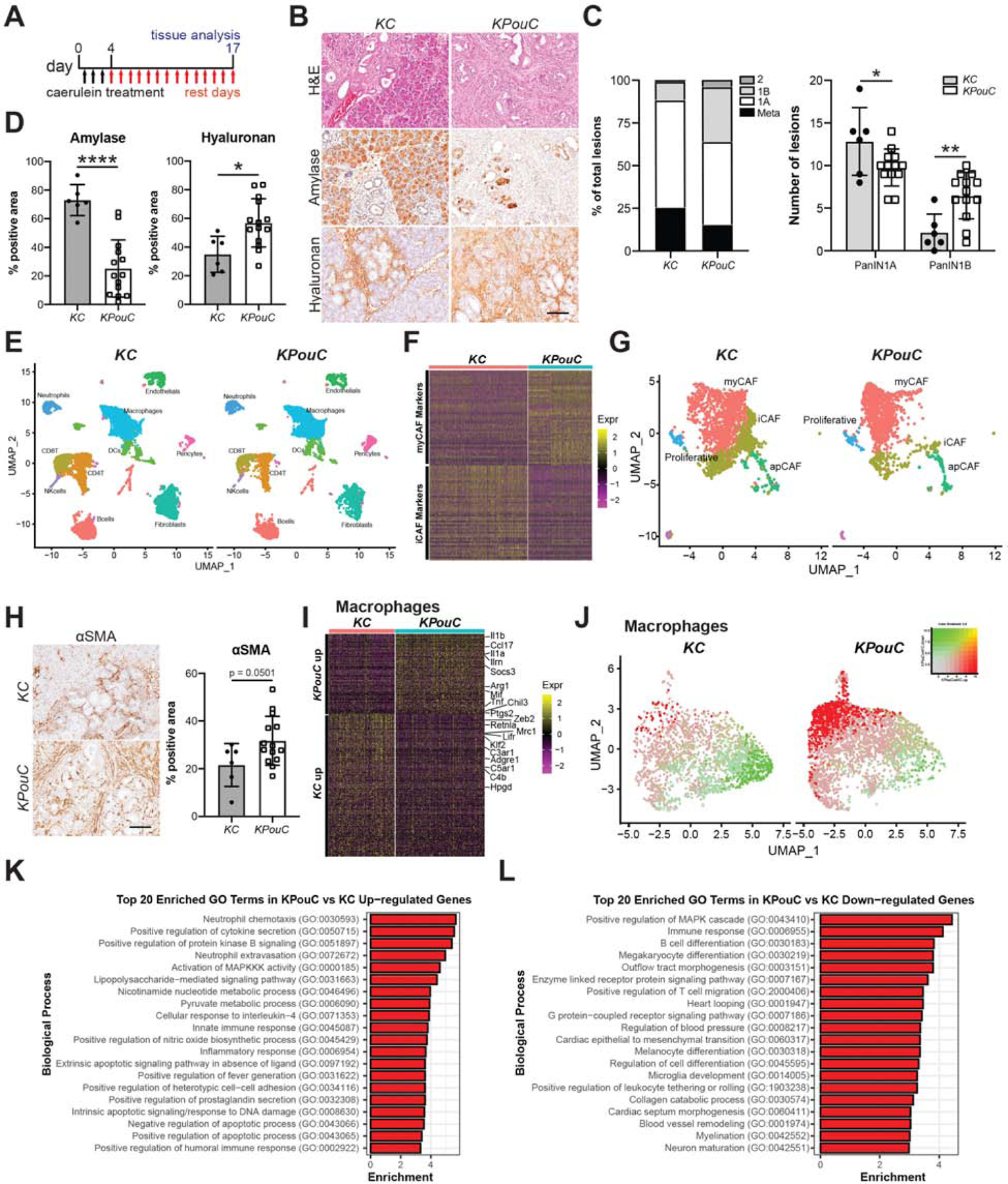
Figure 2. Tuft cell ablation exacerbates pancreatic injury.

(A) Schematic for caerulein treatment of KC and KPouC mice. (B) H&E analysis and immunohistochemistry for acinar cell marker amylase and extracellular matrix component hyaluronan, both brown. (C) PanIN distribution in caerulein-treated KC and KPouC mice. (D) Quantification of histology shown in (B). (E) UMAP showing annotated clusters of stromal populations in injured KC and KPouC pancreata. (F) Heat map showing the top 50 most highly expressed genes described for the myCAF and iCAF subtypes in KC and KPouC fibroblasts. (G) UMAP of fibroblasts from KC and KPouC mice with myCAF, iCAF, apCAF, and proliferative fibroblast RNA signatures overlaid. (H) IHC and quantification for myCAF marker αSMA in KC and KPouC mice. (I) Heat map of the top 100 differentially expressed genes between KPouC and KC macrophages. (J) UMAP of KC and KPouC macrophages overlaid with the module score of KPouC vs. KC up-regulated and down-regulated genes (shown in (I),red and green, respectively). (K) Top 20 enriched gene ontology terms in KPouC up-regulated and (L) down-regulated genes. *, p < 0.05; **, p < 0.01; ****, p < 0.001.
