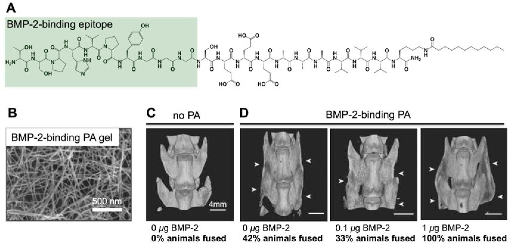
Figure 7.

(A) Chemical structure of a PA containing a short peptide sequence identified to bind BMP-2 via phage display. The bioactive peptide portion is highlighted in green. (B) Scanning electron micrograph (SEM) of a hydrogel containing the BMP-2 binding PA. (C) MicroCT (computerized tomography) reconstruction of an unfused animal treated with no PA and no BMP-2, included for comparison with (D) microCT (computerized tomography) reconstructions of fused animals treated with BMP-2 bindign PA and indicated BMP-2 dosages. The images are specifically of fused animals in the groups; the overall fusion rates164 are indicated. The white arrows indicate the fusion bed. Adapted with permission.164 Copyright 2015, Wiley VCH.
