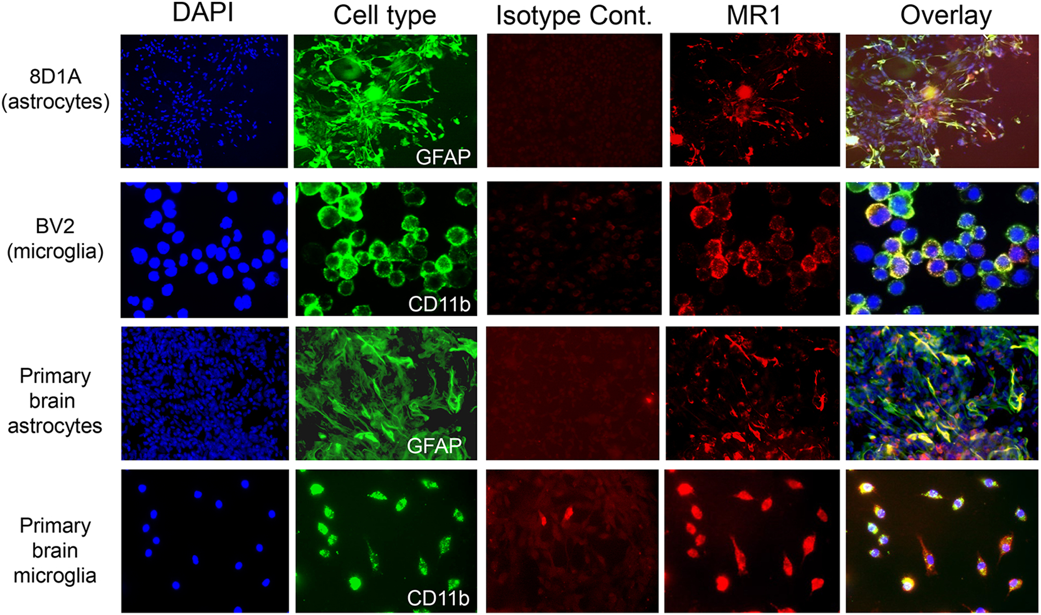
Fig. 2.

Immunofluorescent labelling of mouse brain astrocyte and microglia cell lines and primary cultures. The individual groups of cells were stained with the cell type-specific marker GFAP (astrocytes; green) or CD11b (microglia; green), as well as an anti-MR1 mAb (red) or an isotype control mAb (red). The nuclei were stained with DAPI (blue) and last column shows the overlay for each cell type. Magnification = 20X.
