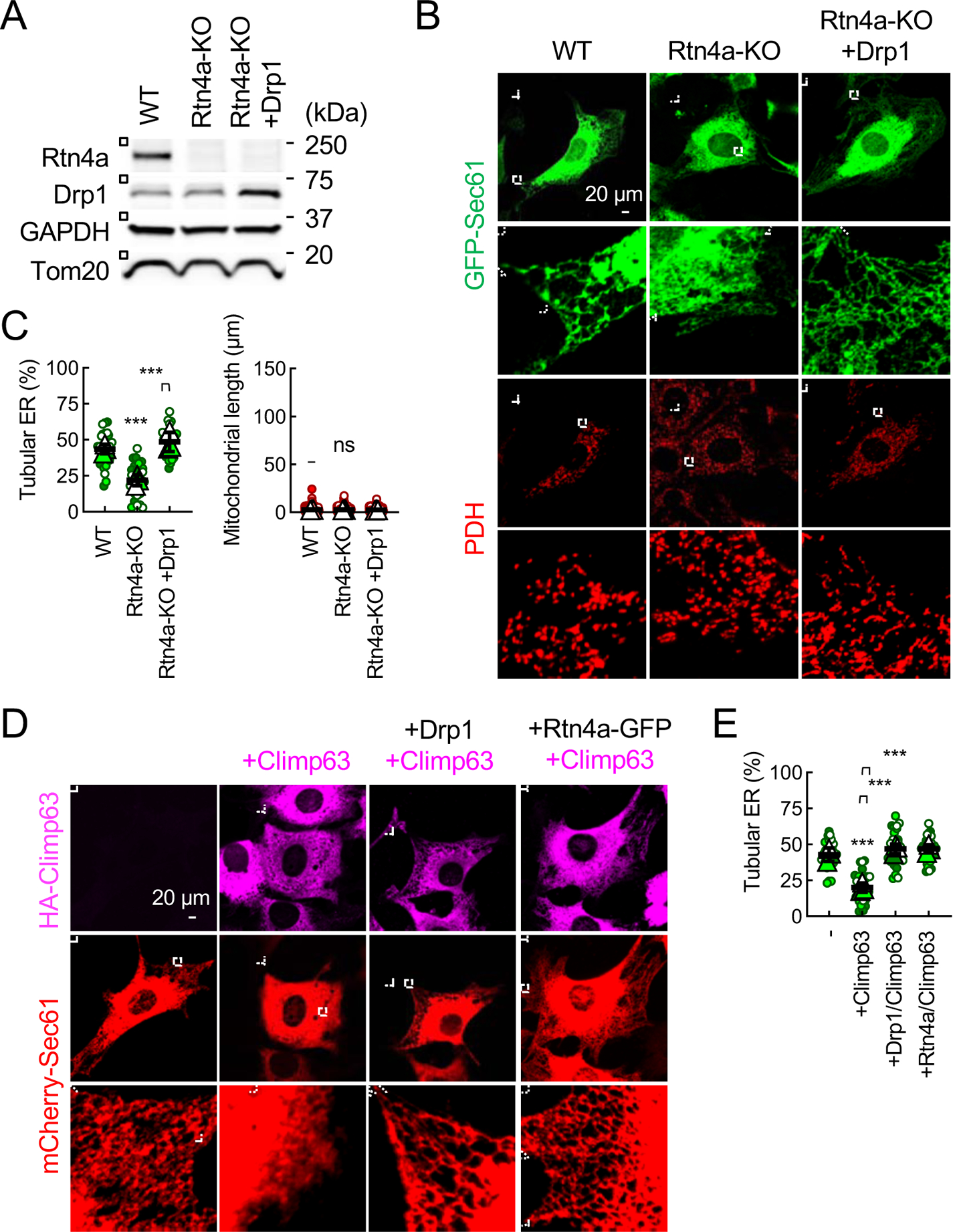
Fig. 3. Drp1 forms ER tubules independently of Rtn4a and Climp63.

(A) The expression levels of Rtn4a and Drp1 were analyzed in WT MEFs, Rtn4a-KO MEFs, and Rtn4a-KO MEFs ectopically expressing Drp1 by Western blotting. (B) The ER and mitochondria in these MEFs were visualized. (C) Quantification of the morphology of the ER and mitochondria. Bars are average ± SD (n = 3 experiments). (D) WT MEF expressing mCherry-Sec61β were transduced with lentiviruses carrying HA-Climp63 along with Drp1 or Rtn4a-GFP. The efficiency of lentivirus transduction was essentially 100%. The MEFs were subjected to immunofluorescence microscopy with antibodies to HA and mCherry. (E) Quantification of ER morphology. Bars are average ± SD (n = 3). Statistical analysis was performed using One-way ANOVA with post-hoc Tukey (C and E): **p<0.01, ***p<0.001.
