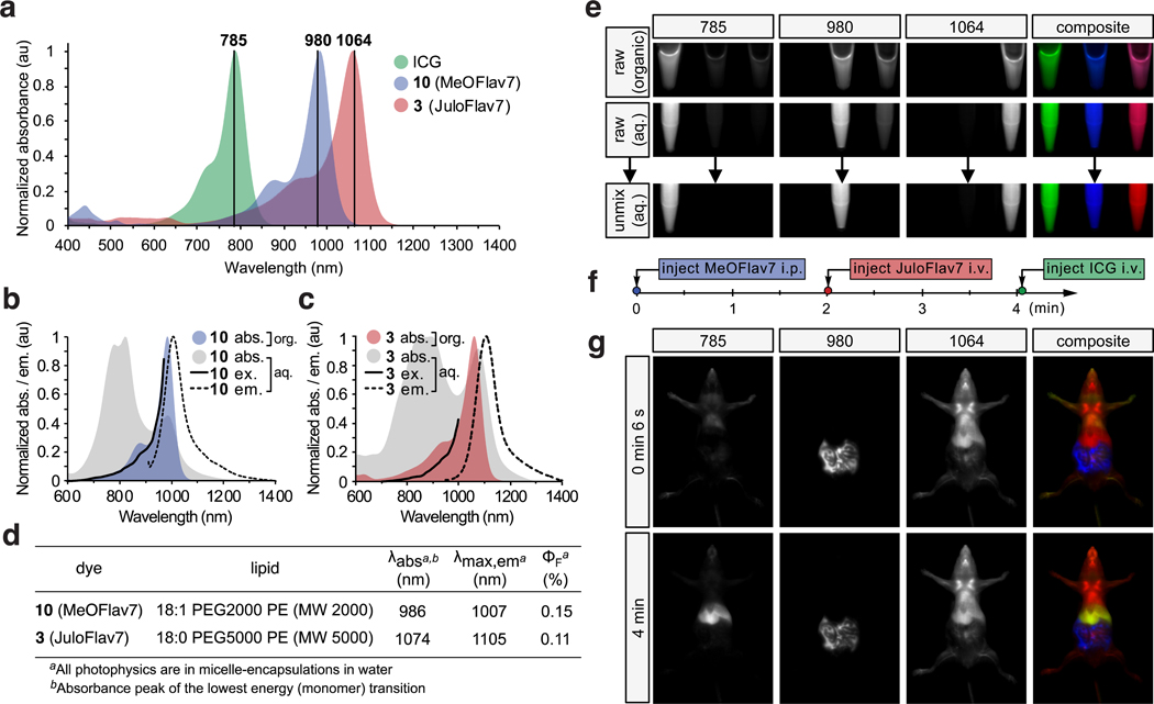
Fig. 3.
Excitation-multiplexed SWIR imaging. a) Absorption profiles of dyes used in imaging experiments plotted against excitation wavelengths employed. b-c) Absorption (grey), emission (ex. at 880 nm (b) and 900 nm (c), black dotted), and excitation spectra (em. monitored at 1008 nm (b), and 1088 nm (c), black solid) of micelle-encapsulated 10 (b) and 3 (c) overlaid with absorption traces of dyes in DCM (colored). d) Photophysics of the micelle-encapsulated dyes in water. e) Raw and unmixed images of successive frames and merged 3-color images of vials containing ICG (left), 10 (center), and 3 (right) in ethanol or DCM (top) and in micelles in water (middle and bottom). Arrow indicates linear unmixing procedure. f) Experimental timeline of administration of the three probes used in (g). g) Multiplexed in vivo images using 785, 980, and 1064 nm ex. (78 mWcm−2) and 1150–1700 nm collection (10 ms exposure time; 27.8 fps). Displayed images are averaged over 5 frames.

