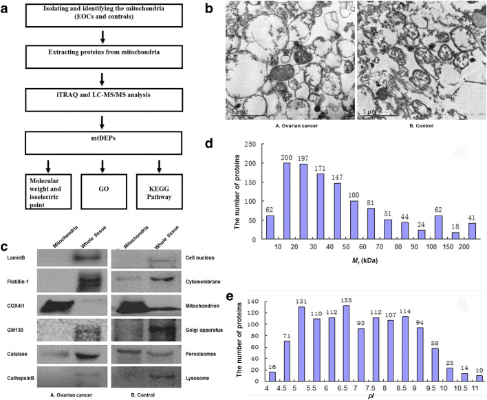
Fig. 1.
Identification of mitochondrial differentially expressed proteins in EOCs relative to controls. a Experimental flowchart to study mitochondrial differentially expressed proteins. b Electron micrograph analysis of mitochondria isolated from epithelial ovarian cancer (A) and control (B) tissues. c Organelle-specific antibody-based western blot analysis of mitochondria isolated from epithelial ovarian cancer (A) and control (B) tissues. Equal amounts of proteins were loaded onto a 10% SDS-PAGE and analyzed by western blotting with indicated antibodies against marker proteins from the cell nucleus, cytomembrane, mitochondrion, Golgi apparatus, peroxisomes, and lysosome. d Distribution status of 1198 mtDEPs according to their molecular mass (Mr). e Distribution status of 1198 mtDEPs according to their isoelectric points (pI). EOC, epithelial ovarian carcinoma; SDS-PAGE, sodium dodecyl sulfate polyacrylamide gel electrophoresis; mtDEPs, mitochondrial differentially expressed proteins; iTRAQ, isobaric tags for relative and absolute quantitation; LC-MS/MS, liquid chromatography-tandem mass spectrometry; GO, Gene Ontology; GM130, golgin A2; KEGG, Kyoto Encyclopedia of Genes and Genomes; COX4I1, cytochrome c oxidase subunit 4I1

