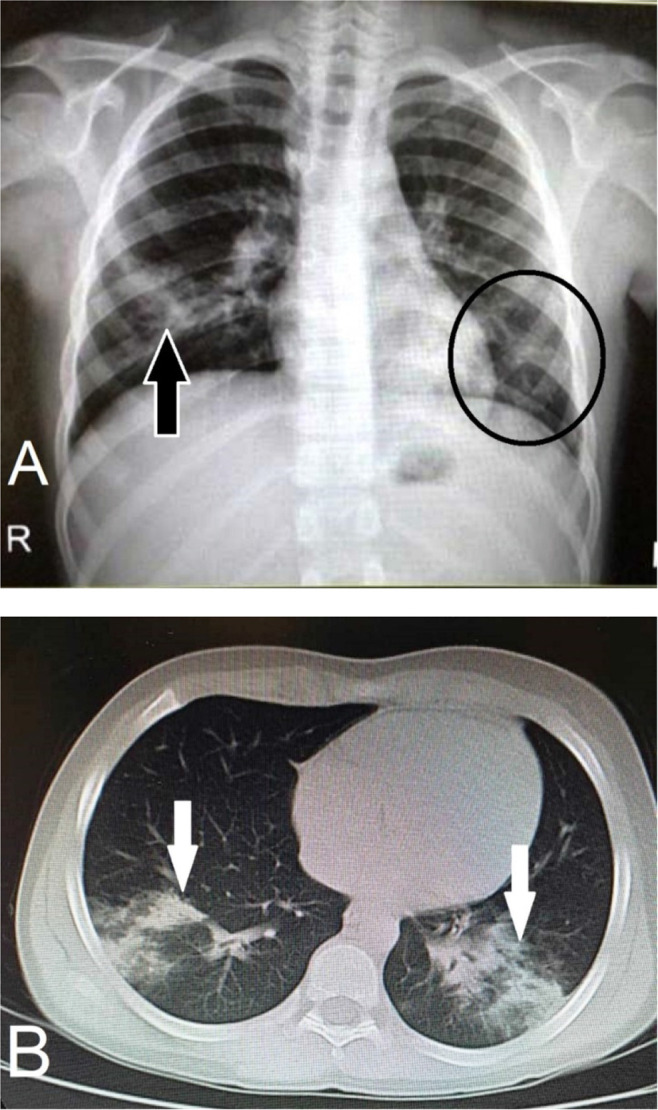
Figure 1.

A) In chest X-ray radiography, air space opacification is visible in the right lower lobe (black arrow), and faint ground-glass opacity can be seen in the left lower lobe (encircled). B) The axial view of chest CT scan for the lungs shows patchy lower lobe consolidaions bilaterally (white arrows).
