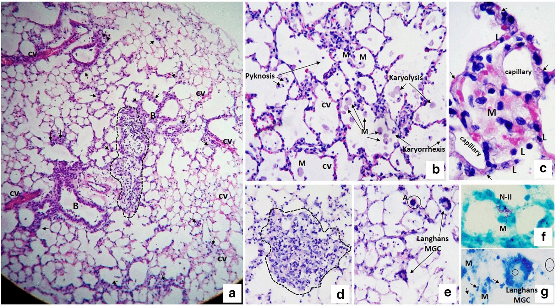
Fig. 2.

Morphological aspects of the infection of murine PCLS infected for 48 h with M. abscessus. a Panoramic view of a complete lung slice infected with M. abscessus for 48 h showing preserved tissue viability and an inflammatory aggregate with a loss of alveolar spaces (dotted area). Vascular congestion (vc) and discrete inflammatory infiltrate in the peribronchioles (B) with septal thickening (arrowhead) are also observed. b Alveolar spaces with an increase in foamy macrophages (FM), some of which show nuclear changes such as pyknosis, karyolysis and karyorrhexis. In the alveolar septa, vascular congestion and moderate inflammatory infiltrate are observed. c The alveolar space is occupied entirely by a conglomerate of foamy macrophages mixed with erythrocytes; the alveolar septa show discrete thickening secondary to edema, vascular congestion (arrows) and lymphocyte infiltrate (L). In addition, normal capillary vessels are observed adjacent to the septa. d, e Photomicrograph showing lung tissue with a loss of its histological structure and occupied by inflammatory infiltrates of mononuclear cells, polymorphonuclear cells, and histiocyte/macrophages (dotted area). The rest of the tissue shows septal inflammation and vascular congestion. In addition to the histological findings in the photomicrograph (c), multinucleated giant Langhans cells and apoptotic cells (A) are observed. f Abundant mycobacteria in the alveolar septum infecting type II pneumocytes (N-II) and foamy macrophages (FM). g Mycobacterial fragments (circles) are observed within Langhans multinucleated giant cell and extracellularly. Foamy macrophages (FM) and nuclear fragments (arrows) can also be observed. H&E staining (a–e). ZN staining (f, g). Total magnification: ×50 (a), ×100 (d, e), ×200 (b), ×400 (c, f, g)
