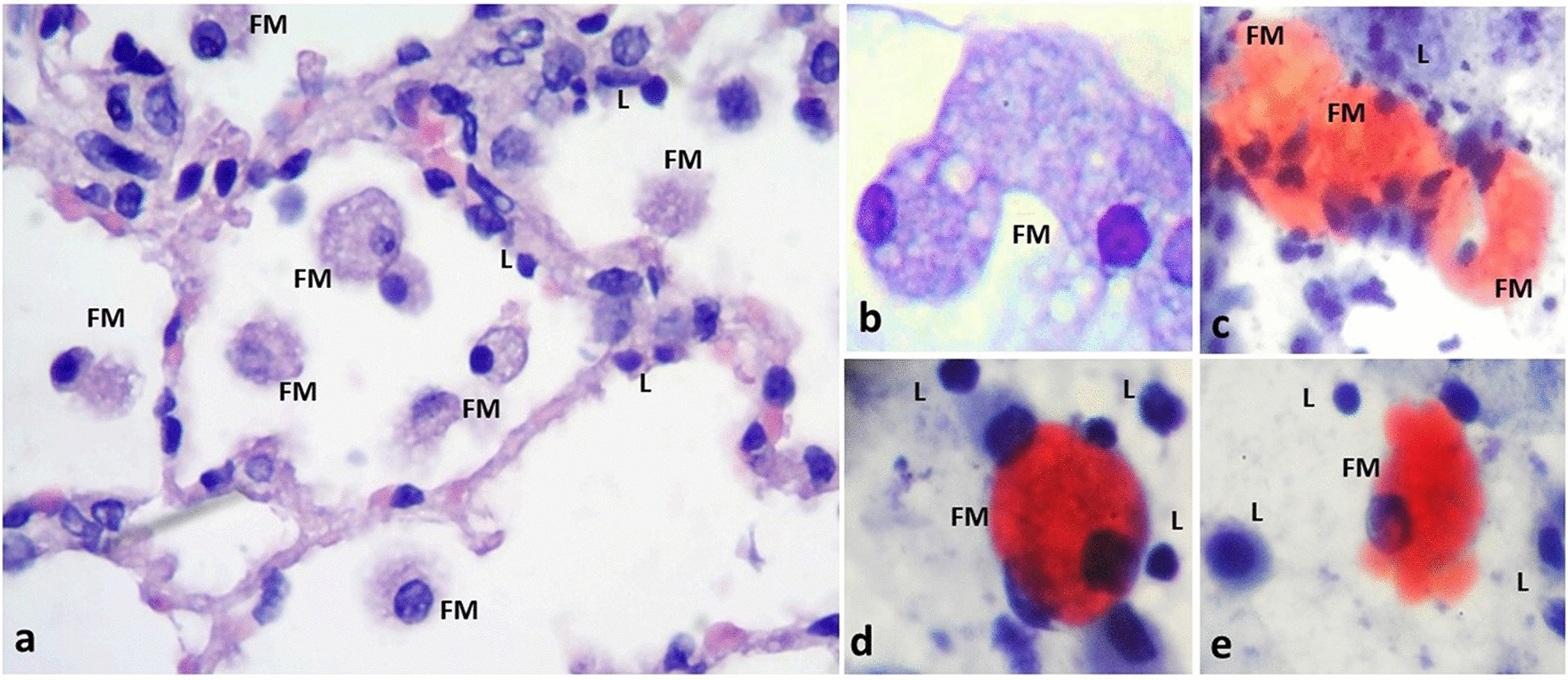
Fig. 4.

Presence of foamy macrophages in the infected PCLS. Inflammatory infiltrates with aggregates of foamy macrophages were frequently observed in the PCLS infected with M. abscessus. a Alveolar spaces with abundant foamy macrophages (FM). b Cellular detail of FM containing multiple intracytoplasmic vacuoles. Foamy macrophages (FM) can be found in aggregates (c), or isolated (d, e), with positive staining for accumulation of lipids in their cytoplasm; lipid-laden cells are a distinctive hallmark of mycobacterial infections. Mononuclear cells, predominantly lymphocyes (L) are also observed. H&E staining (a, b). Oil red-O staining (c–e). Total magnification: ×10 (a), ×40 (b–e)
