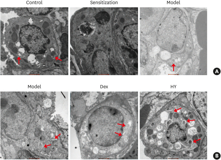
Fig. 4. Effect of HY peptide treatment on AVs in lung tissues. (A) AVs mainly located in ATII, as well as the formation of AVs in lung tissues of asthmatic mice, were decreased under TEM. (B) Electron microscopic examination of AVs after treatment with HY peptide in lung tissues. The red arrows indicate the presence of AVs (scale bar = 1 µm).
AV, autophagic vacuole; Control, control group; Sensitization, sensitization group; Model, model group; Dex, dexamethasone-treated group; HY, HY peptide-treated group; TEM, transmission electron microscopy.

