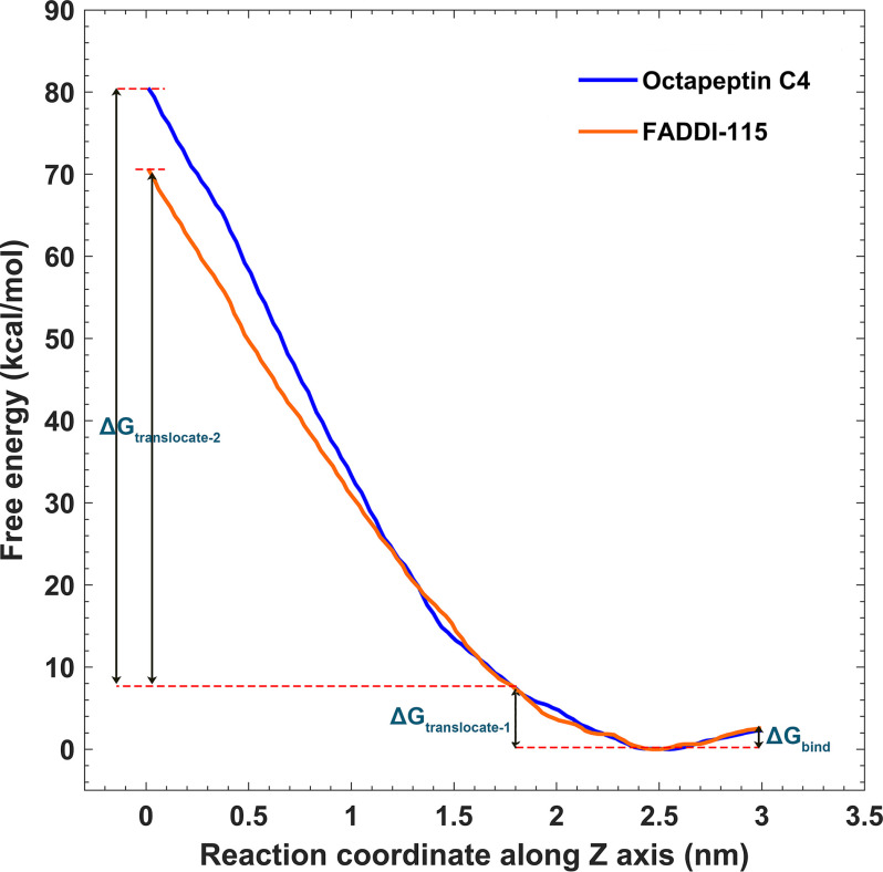
Figure 4.
Free energy profiles for the penetration of octapeptin C4 and FADDI-115 into the outer membrane. The free energy barriers for octapeptin binding to the outer membrane, passing through the headgroup and hydrocarbon regions of the outer leaflet are depicted by ΔGbind, ΔGtranslocate-1, and ΔGtranslocate-2, respectively. The reaction coordinates along z axis represent the positions relative to the center of the outer membrane, with Z = 0 indicating the hydrophobic center of the outer membrane.

