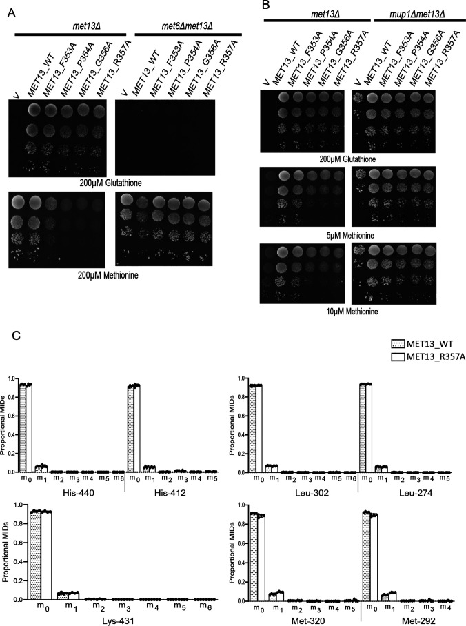
Figure 7.
Methionine biosynthesis continues in the deregulated MTHFR even in the presence of exogenous methionine. Yeast met13Δ and met6Δmet13Δ (A) or mup1Δmet13Δ (B) strains were transformed with vector control (V), MET13_WT, MET13_F353A, MET13_P354A, MET13_G356A, and MET13_R357A were grown to exponential phase in minimal medium containing 200 μm GSH, harvested, washed, resuspended in water, and serially diluted to give 0.1, 0.01, 0.001, and 0.0001 A600 of cells. 10 μl of these dilutions were spotted on minimal medium containing 200 μm GSH or different concentrations of methionine. The photographs were taken after 48 h of incubation at 30 °C. The experiment was repeated three times, and a representative data set is shown. C, 13C label redistribution, fate of externally fed, histidine, lysine, leucine, and methionine is depicted. The proportional MIDs of protein-derived amino acid fragments retrobiosynthetically report on the labeling of precursors. These amino acid fragments were obtained by TBDMS derivatization and GC-MS ionization and are presented by their m/z and carbon backbone.

