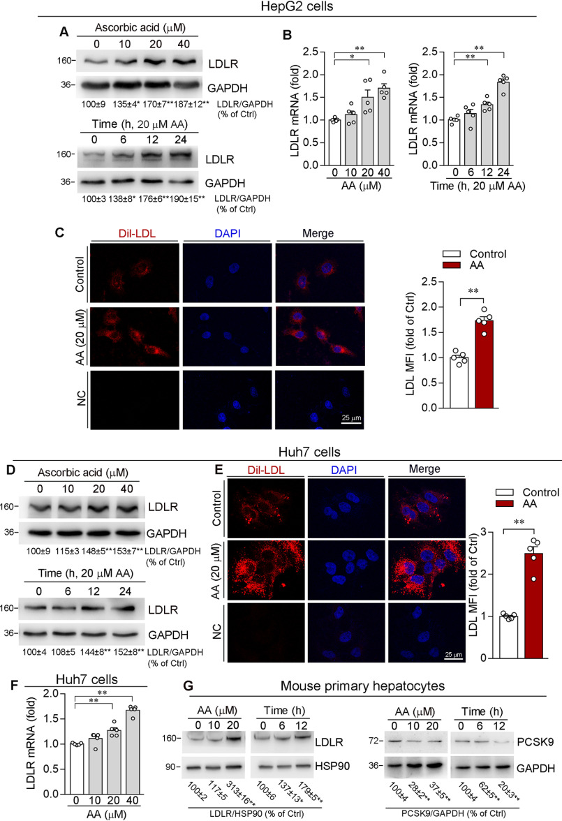
Figure 2.
Ascorbic acid enhances LDLR expression and LDL uptake by hepatic cell lines. HepG2 or Huh7 cells were treated with AA at the indicated concentrations for 12 h or with 20 μm AA for the indicated times. LDLR protein or mRNA expression was determined by Western blotting (A and D) or qRT-PCR (B and F). C and E, HepG2 or Huh7 cells were treated with 20 μm AA for 12 h. Cells were then switched to serum-free medium containing Dil-LDL (20 µg/ml) and incubated for 4 h at 37 °C, followed by photograph with a fluorescence microscope. The MFI in images was quantitatively analyzed (original magnification, ×400). G, primary hepatocytes isolated from C57BL/6J mice were treated with AA at the indicated concentrations for 12 h or with 20 μm AA for the indicated times. Expression of PCSK9 or LDLR protein was determined by Western blotting. *, p < 0.05; **, p < 0.01 versus control (n = 5).

