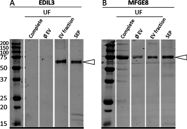
Figure 5.

Western blotting analysis of (A) EDIL3 and (B) MFGE8 in UF: unfractioned UF (complete), depleted UF (ØEV), EV fraction of UF (EV fraction), and SEP. Proteins (15 µg) were subjected to 12.5% SDS-PAGE electrophoresis and blotted for analysis. The membranes were probed with (A) rabbit polyclonal anti-EDIL3 (1:1,000, SAB2105802, Sigma-Aldrich, Saint-Quentin Fallavier, France) and (B) rabbit synthetic anti-MFGE8 peptide (1:1,000, rabbit polyclonal; ProteoGenix, Schiltigheim). White arrowheads indicate the immunoreactive bands.
